Science
 BRAIN NETWORKS IN MENTAL ILLNESS
A new way of analyzing brain scans is yielding extraordinary new insights.
by John McManamy
A new way of analyzing brain scans is yielding extraordinary new insights.
by John McManamy
RESTING STATE FUNCTIONAL CONNECTIVITY - savor the resonance. We know, for instance, the amygdala doesn't operate in isolation, that it is networked to other areas of the brain. But in what ways? We are used to seeing what happens when the brain is activated, but figuring out the brain in its resting state may tell us more.
The old way of mapping brains in their resting states was to zero in on a target area such as the amygdala and try to pick up what other signals from elsewhere may have been occurring at the same time. According to Dr Volkow (with Dardo Tomasi of the National Institute on Alcohol Abuse and Alcoholism), in an article in PNAS:
More recently researchers have started to use data-driven approaches that are based on graph theory to assess the functional connectivity of the human brain using datasets obtained with MRI.
I have no idea what this means, but the bottom line is the new way is 1000 times faster than old techniques, which suddenly makes mapping functional connectivity a doable exercise.
In her talk, Dr Volkow described scanning brains in a resting conscious state, paying attention to oscillations - generally regarded as background noise - across different regions. Oscillations occurring at the same time could be assumed to be part of the same functioning network. To test this, Volkow and Tomasi analyzed the brain scans of 979 subjects from 35 centers around the world. One of the things they were looking for was "local functional connectivity density."
Brain hubs, to put it into layman's terms. In the authors' words:
Brain networks with energy-efficient hubs might support the high cognitive performance of humans and a better understanding of their organization is likely of relevance for studying not only brain development and plasticity but also neuropsychiatric disorders.
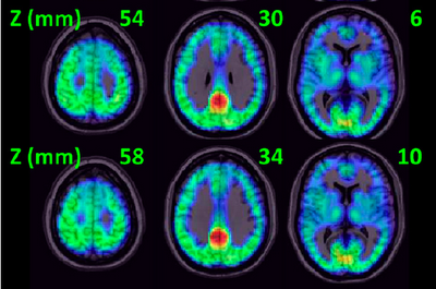
Volkow and Tomasi's analysis located the mother of all hubs in the posterior cingulate, corresponding with Brodman Area 23. The cingulate snakes beneath the outer cortices and, among many things, plays a major role in mediating back and forth traffic between the limbic system and thinking parts of the brain. We have known for some time that the cingulate acts as a major hub, but if I am interpreting the authors correctly, we are talking Grand Central Station dimensions.
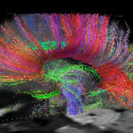
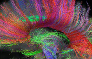
In the image below, the red-orange indicates the highest signal strength, corresponding with the posterior cingulate/ventral precuneus.


SIGN UP FOR MY FREE EMAIL NEWSLETTER
The very strong implication is that things going smoothly in the cingulate bode well for things going smoothly in the rest of the brain. In her talk, Dr Volkow mentioned deep brain stimulation (DBS) for depression, focusing on Brodman Area 25, in the immediate hub neighborhood. DBS is also being applied to other brain regions that appear linked to BA 25. Food for thought.
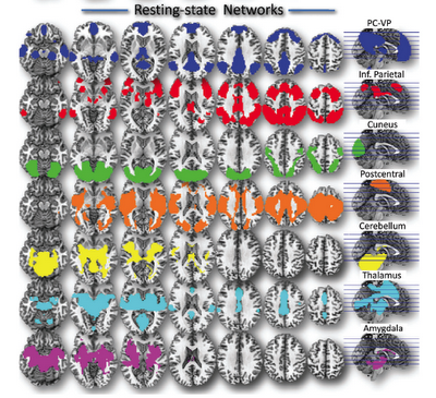
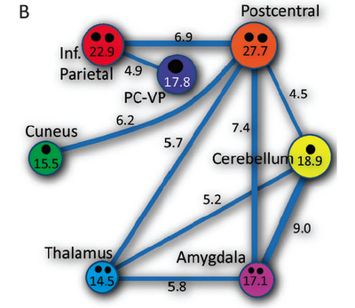
From their hub findings, Volkow and Tomasi identified seven overlapping networks covering 80 percent of all the gray matter in the brain. These involved four major cortical networks (default mode, dorsal attention, visual, and somatosensory) linked to four hubs (ventral precuneus/posterior cingulate, inferior parietal cortex, cuneus, and postcentral gyrus), and three subcortical (tied to hubs involving the cerebellum, thalamus, and amygdala).
The authors note that to keep things simple, they kept the hub count low, based on signal strength and volume. I think this means that researchers will be kept busy for a long time mapping out minor hubs. But the low hub count confirms something that experts have known for a long time, namely that all manner of illnesses and conditions share many of the same pathways. Indeed, Dr Volker in her talk pointed out that both drug use and stress increase dopamine activity in the nucleus accumbens.
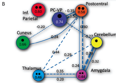
Below are renderings of the different networks. (Note, postcentral roughly corresponds to the posterior cingulate.)


This is what they look like together:
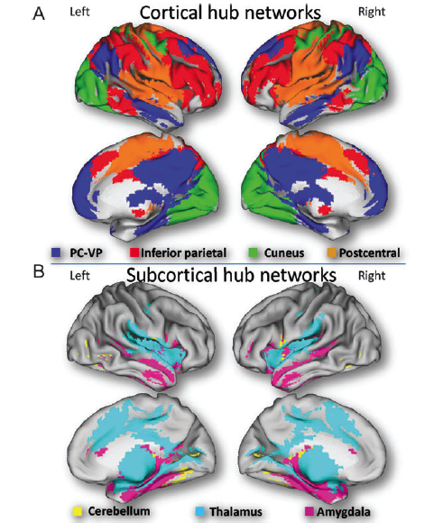
Here is a simple diagram illustrating the overlap:
Here is a more complex one:
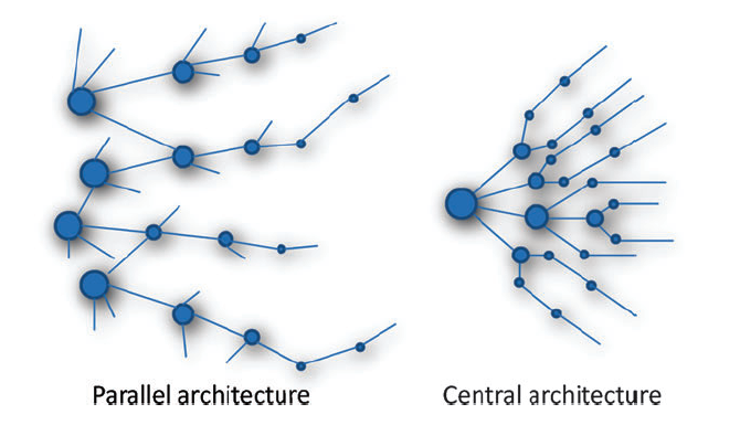
In her talk, Dr Volkow indicated that the evidence points to the brain organized according to parallel architecture rather than central, sort of like this:
The field of resting state functional connectivity is so new tback when I first published this article in 2012, Wikipedia did not have an entry for it. Don't worry. It does now..
Reviewed July 13, 2016
NEW!
Follow me on the road. Check out my New Heart, New Start blog.